Clinical Evaluation
Risk Factors
The different types of allergic conjunctivitis with corneal involvement have different risk factors.3
- Vernal Keratoconjunctivitis (VKC): This is a more severe form of allergic conjunctivitis, typically seen in children and young adults. It is rarer than SAC and PAC, but due to possible corneal involvement, it is potentially a more serious condition. VKC has a mean age of onset of 7 years and the majority of cases (85%) have an onset prior to age 10 years.4 It is found most commonly in males.5
- Atopic Keratoconjunctivitis (AKC): AKC is a chronic and severe form of allergic conjunctivitis often associated with atopic dermatitis (eczema)6 and other systemic allergies. It is the most potentially blinding of all the ocular allergies. Similar toVKC, AKC is more common in males. The condition typically begins in the late teens or early twenties with a peak severity occurring between ages 30 to 50 years.6
Signs
The different types of allergic conjunctivitis have different presenting signs.
Vernal Keratoconjunctivitis:3, 7
- Discharge that is stringy white and mucous
- Palbebral
- Hyperaemia and chemosis of the conjunctiva
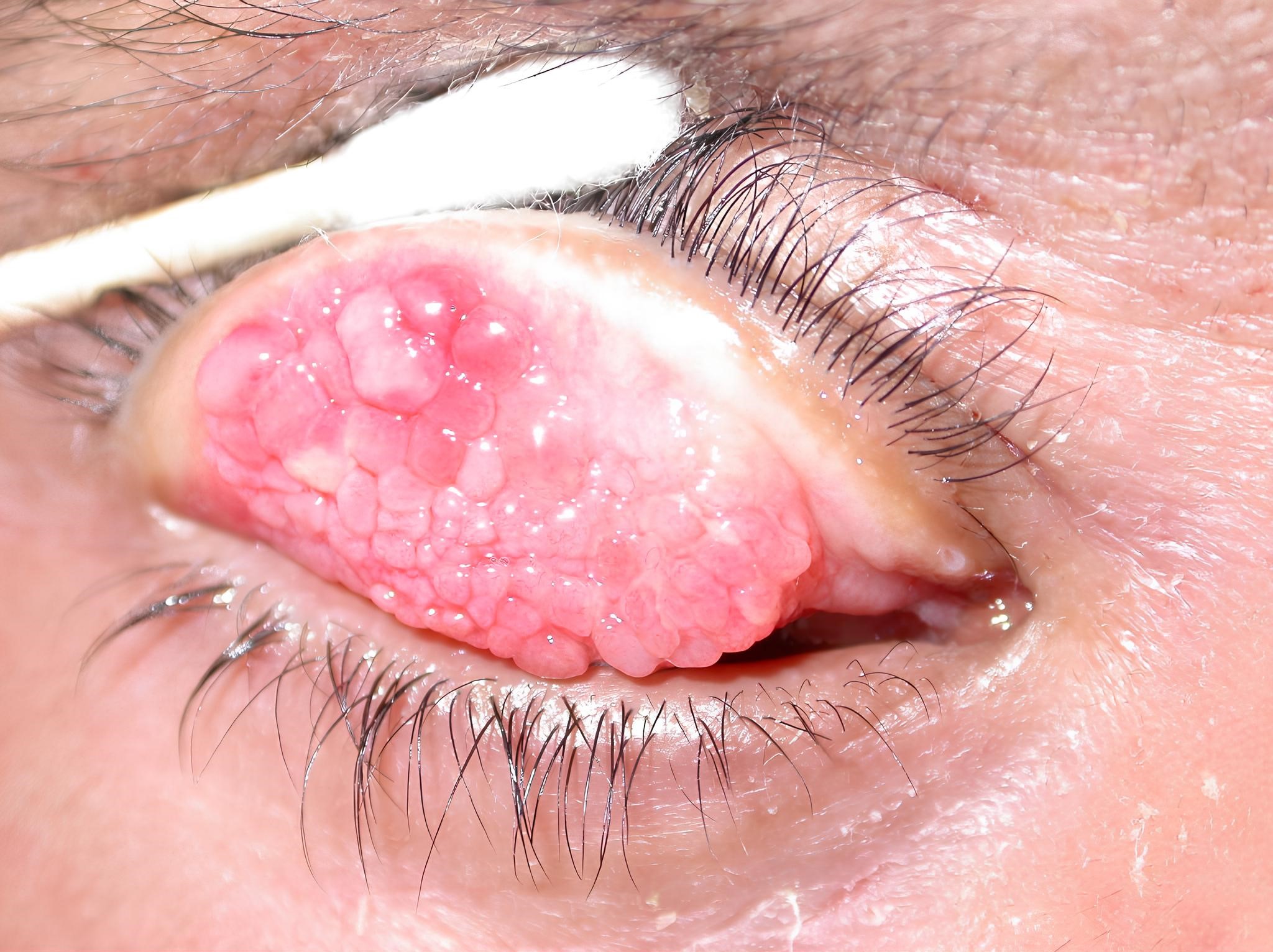
- Giant tarsal papillae
- Limbal
- Hyperaemia and oedema causing thickening
- Trantas Dots (small white/yellow accumulations of eosinophils which may be unilateral)
- Corneal (Usually upper third)
- Punctate epithelial keratopathy
- Macro-erosions
- Plaque or shield ulcer
- Subepithelial scarring
- Unlike SAC and PAC the signs may be different from one eye to the other
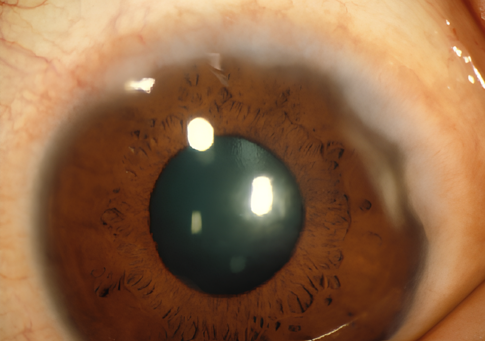
- Patients with VKC may also have keratoconus and atopic cataract (anterior or posterior polar)
Atopic Keratoconjunctivitis:3, 8
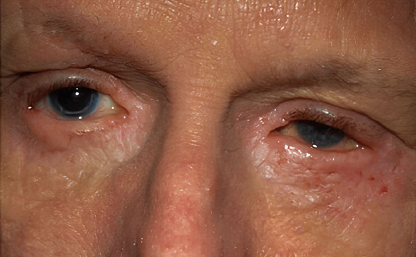
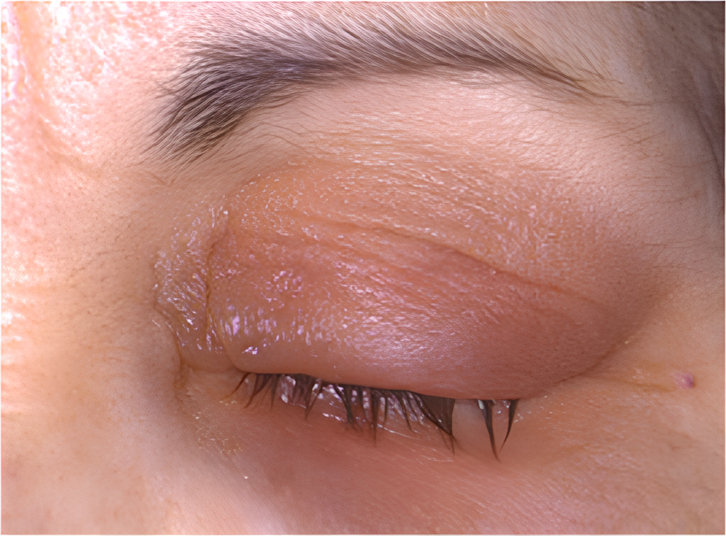
- Thickened eyelids with crusting
- Blepharitis (staphylococcal)
- Conjunctival hyperaemia
- Giant papillary hypertrophy
- Limbal inflammation
- Punctate corneal epitheliopathy which may progress to macro erosion, plaque formation, subepithelial scarring, neovascularisation, pannus, thinning and possible spontaneous perforation.
- There is a greater likelihood that these patients develop herpes simplex keratitis, keratoconus, atopic cataract, and retinal detachment.
Symptoms
The different types of allergic conjunctivitis will have different presenting symptoms, but one of the most important key factors in ocular allergy is itch.
Vernal Keratoconjunctivitis:3, 7
- Itchy, burning, or a sensation of a foreign object in the eyes
- Excessive tearing
- Presence of stringy, mucus-like discharge
- Vision may become blurred
- Pain may occur if the cornea is affected
- Sensitivity to light, sometimes severe
- Difficulty opening the eyes upon waking
- Note: Symptoms commonly affect both eyes, though often with asymmetry in severity
Atopic Keratoconjunctivitis:3, 8
- Itching, burning, and watering in the eyes – typically experienced in both eyes
- Blurred vision and sensitivity to light may also be present
- A white, stringy, mucoid discharge from the eyes is common
- Ocular symptoms may start several years after the onset of atopic conditions
- These symptoms usually affect both eyes, persist throughout the year, and may worsen periodically
Investigations
History
A clinical history will help clinicians to differentiate ocular allergy from other potential eye conditions.3 Nearly 49% of patients who suffer from VKC have a family history of atopic disease such as asthma, rhinitis, or eczema. VKC is also commonly associated with a personal allergy history, which might include asthma, rhinitis, or atopic eczema. Therefore, it’s important to check for signs of eczema and other atopic symptoms during the examination of an AKC patients. As for example, distinguishing systemic manifestations of atopic dermatitis can aid in distinguishing AKC from VKC.
Slit Lamp Examination:3
This will help clinicians to differentiate ocular allergy from other potentially sight threatening ocular conditions, and the following may be considered in examining these patients:
- Evaluate lids and lashes to look for swelling, thickening, signs of eczema, and blepharitis.
- Look for conjunctival chemosis
- Check and grade conjunctival redness.
- Look for limbal changes.
- Evert eyelids to look for and grade papillae and hyperaemia
- Check cornea (No corneal involvement in acute allergic, seasonal, perennial or giant papillary conjunctivitis)
- Check anterior chamber
- Look for atopic cataract
- Check for possible retinal detachment
Diagnostic Methods
- Skin Testing: Utilising a skin-prick test, which has been reported to demonstrate positivity in only 20% of patients with ocular allergies.9
- Conjunctival Provocation Test: Involves the application of allergens topically into the conjunctival sac to elicit a response.10
- Biopsy of Conjunctiva: Involves obtaining cytology specimens from the conjunctiva, with a focus on identifying the presence of eosinophils and mast cells. The presence of eosinophils during an allergic reaction is recognised as diagnostic for allergy.10
- Tear Fluid Analysis: A measurement of IgE levels greater than 16 IU/ml in tear fluid can be indicative of allergic conjunctivitis. However, tear analysis does not differentiate between different types of ocular allergies.10