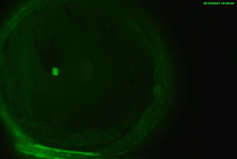
Courtesy of Mr Sathish Srinivasan – Consultant Corneal Surgeon, Joint Clinical Director, University Hospital Ayr, Scotland, UK and Director of Ayrshire Eye Clinic
Clinical Images & Videos
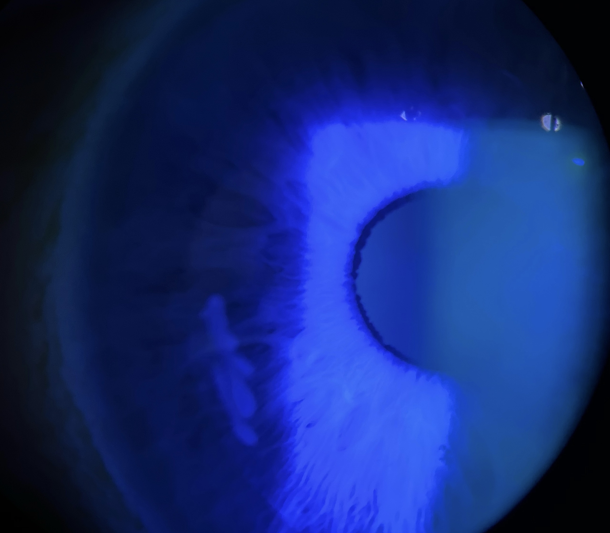
Epithelial ingrowth fluorescein
Share Epithelial ingrowth fluorescein via QR code
Please instruct your patient or colleague to scan the QR code with the camera app on their smartphone to access this resource.
They can scan the QR code directly from your device if they are there with you in person.
Alternatively you can also copy and past the QR code into an email or document with additional information that you want to provide to them.
Photos
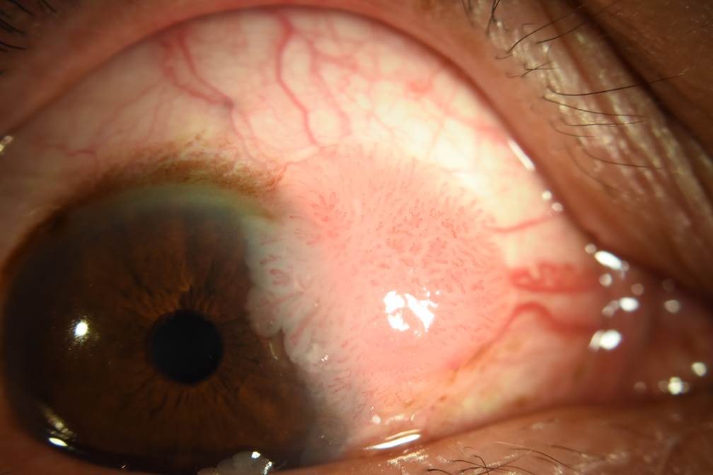
Conjunctival papilloma
Courtesy of Mr Sathish Srinivasan – Consultant Corneal Surgeon, Joint Clinical Director, University Hospital Ayr, Scotland, UK and Director of Ayrshire Eye Clinic
Share Conjunctival papilloma via QR code
Please instruct your patient or colleague to scan the QR code with the camera app on their smartphone to access this resource.
They can scan the QR code directly from your device if they are there with you in person.
Alternatively you can also copy and past the QR code into an email or document with additional information that you want to provide to them.
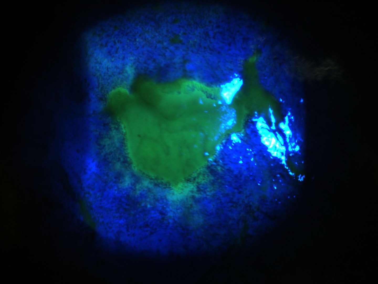
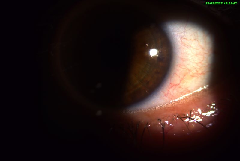
Central persitant corneal epi defect due to 360 degree limbal stem cell failure
Courtesy of Mr Sathish Srinivasan – Consultant Corneal Surgeon, Joint Clinical Director, University Hospital Ayr, Scotland, UK and Director of Ayrshire Eye Clinic
Share Central persitant corneal epi defect due to 360 degree limbal stem cell failure via QR code
Please instruct your patient or colleague to scan the QR code with the camera app on their smartphone to access this resource.
They can scan the QR code directly from your device if they are there with you in person.
Alternatively you can also copy and past the QR code into an email or document with additional information that you want to provide to them.
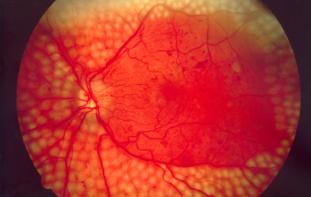
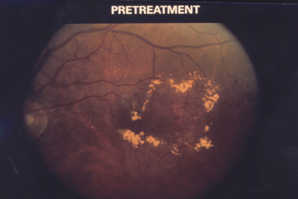
Fundus photo showing scatter laser surgery for diabetic retinopathy.
Courtesy of the National Eye Institute/National Institutes of Health available at https://medialibrary.nei.nih.gov/
Share Fundus photo showing scatter laser surgery for diabetic retinopathy. via QR code
Please instruct your patient or colleague to scan the QR code with the camera app on their smartphone to access this resource.
They can scan the QR code directly from your device if they are there with you in person.
Alternatively you can also copy and past the QR code into an email or document with additional information that you want to provide to them.
Photos
Diabetic Macular Edema
Diabetic Macular Edema – Courtesy of the National Eye Institute/National Institutes of Health available at https://medialibrary.nei.nih.gov/
Share Diabetic Macular Edema via QR code
Please instruct your patient or colleague to scan the QR code with the camera app on their smartphone to access this resource.
They can scan the QR code directly from your device if they are there with you in person.
Alternatively you can also copy and past the QR code into an email or document with additional information that you want to provide to them.
Photos
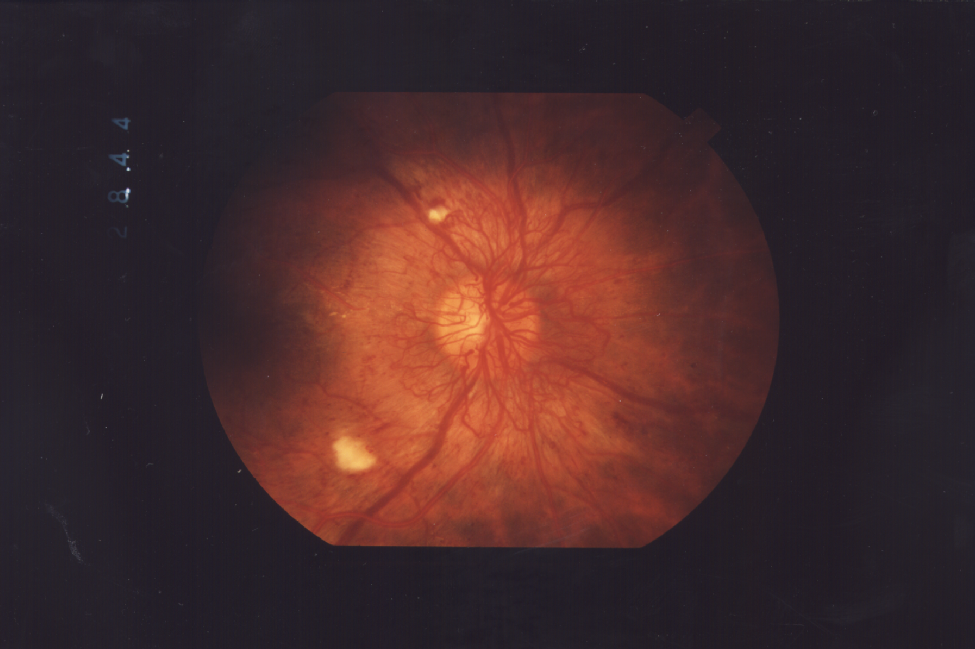
Proliferative Retinopathy
Proliferative retinopathy, an advanced form of diabetic retinopathy, occurs when abnormal new blood vessels and scar tissue form on the surface of the retina – Courtesy of the National Eye Institute/National Institutes of Health available at https://medialibrary.nei.nih.gov/
Share Proliferative Retinopathy via QR code
Please instruct your patient or colleague to scan the QR code with the camera app on their smartphone to access this resource.
They can scan the QR code directly from your device if they are there with you in person.
Alternatively you can also copy and past the QR code into an email or document with additional information that you want to provide to them.
Photos
Share Frothy Tears via QR code
Please instruct your patient or colleague to scan the QR code with the camera app on their smartphone to access this resource.
They can scan the QR code directly from your device if they are there with you in person.
Alternatively you can also copy and past the QR code into an email or document with additional information that you want to provide to them.
Photos
Share Blocked Meibomian Gland via QR code
Please instruct your patient or colleague to scan the QR code with the camera app on their smartphone to access this resource.
They can scan the QR code directly from your device if they are there with you in person.
Alternatively you can also copy and past the QR code into an email or document with additional information that you want to provide to them.
Photos

Share Inferior Corneal Stain via QR code
Please instruct your patient or colleague to scan the QR code with the camera app on their smartphone to access this resource.
They can scan the QR code directly from your device if they are there with you in person.
Alternatively you can also copy and past the QR code into an email or document with additional information that you want to provide to them.
Photos
Share Inferior Drying via QR code
Please instruct your patient or colleague to scan the QR code with the camera app on their smartphone to access this resource.
They can scan the QR code directly from your device if they are there with you in person.
Alternatively you can also copy and past the QR code into an email or document with additional information that you want to provide to them.