Management & Advice
There is no gold standard treatment for meibomian gland dysfunction, but rather a diversity of options.1 The goal of all MGD treatments is to improve the flow of the meibum, leading to a more stable tear film.6 There is a range of treatment options available for the management of MGD that are non-pharmcological treatments including warm compresses, cleansing agents, lubricant drops, and essential fatty acid supplementation; medical treatments including antibiotics, non-steroidal and steroidal anti-inflammatory agents, hormone therapy; control of Demodex infestation and procedures including, gland expression, intraductal meibomian gland probing, the use of electronic heating devices, intense pulsed light therapy, and intranasal neurostimulation.
Non-Pharmacological Treatments
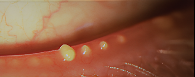
Warm Compresses: Due to many factors as listed above the meibum may be of poor quality or obstructed. It is therefore necessary to use heat to liquify the meibum to allow it to be expressed more freely. Evidence suggests that the melting point of meibum is between 32°C-45°C,2 the poorer the quality, or the more the obstruction the higher the melting point will be within the range of 32°C-45°C.2
Older versions of the warm compress would be a warm face cloth over the eyelids; however, we now know that this would mean the heat would dissipate very quickly and to improve the quality of the meibum you need consistent heat of a temperature of ≥40°C.8,9 Studies have shown that comparing a warm face cloth and a heat mask, a face cloth has no effect on the MGD however, the heat mask reduces the MGD. 8
After the heat is applied, it is recommended to express the glands by form of gentle massage.7 The international workshop on MGD recommends that “patients may be told that after application of a hot compress to the eyelids, they should apply traction on the lateral canthus to immobilize the upper and lower eyelids; that should be followed by down- or upward mild compression of the eyelids with the finger of the opposite hand beginning at the nasal canthus and moving laterally toward the lateral canthus”.7
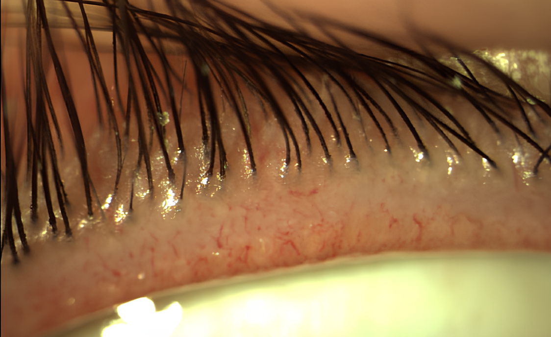
Eyelid Hygiene: Because of the close proximity of the eyelashes to the meibomian glands disorders of one can affect both.9 It has been reported that ecto-parasites demodex folliculorum (DF) tend to live in the eyelash follicles and demodex brevis (DB) tend to burrow into the meibomian glands and are associated with MGD. 9 Treatment is needed to reduce the quantity of DB and DF which has shown to improve patient symptoms and ocular surface health. 9
It has been found that high heat especially ≥40°C causes protein coagulation and denaturation and paralysis of the DF nervous system. This is where warm compresses are useful. It is useful to note that heat masks come in a moist or a dry heat. It has been reported that moist heat helps to soften the eyelash debris in anterior blepharitis.9
It is recommended that patients use an eyelid cleanser (e.g. eyelid wipes, eyelid gel or cleansing solution) to remove debris and create a clean periocular environment.
Lubrication: Preservative free eye drops have shown greater effectiveness than preserved drops in decreasing inflammation on the ocular surface and increasing the antioxidant contents in tears of patients with DED.10 Drops should be recommended at the end of the regime to restabilise the tear film. There are specific drops that can help address the compromised lipid layer by adding lipids to the ocular surface or by helping the lipids that are present work more effectively to create a working lipid layer.
Omega 3 Supplementation: Omega-3 fatty acid supplementation is a potential management option for MGD due to its anti-inflammatory properties and influence on meibum health.13
- Anti-Inflammatory Effects: Omega-3 fatty acids reduce inflammation in the eyelids, addressing the root cause of MGD by improving the function of the meibomian glands, which produce the oily component of tears.
- Modulation of Meibum Composition: Omega-3s may alter the composition of meibum, making it less viscous and more fluid. This modulation prevents blockages in the meibomian glands, enhancing tear quality and reducing symptoms.
- Stabilization of Tear Film: By improving the quality of meibum, omega-3 supplementation stabilizes the tear film, preventing excessive tear evaporation and reducing symptoms of dry eye.
- Reduction of Dry Eye Symptoms: Omega-3 supplements have been shown to alleviate symptoms of dry eye, including dryness, burning, and blurred vision, commonly associated with MGD.
- Potential Improvement in Gland Function: Some studies suggest that omega-3 supplementation may enhance the function of meibomian glands over time, providing long-term benefits for managing MGD and associated dry eye symptoms.
Pharmacological Treatments
In some cases, topical ophthalmic medications such as corticosteroids or antibiotics (eg azithromycin) may be considered. Azithromycin has anti-inflammatory effects and has been shown to improve signs of eyelid disease. Weak topical corticosteroids may also be prescribed and while these drugs don’t directly act on the meibomian glands, they do reduce eyelid inflammation. A short course of these is to be considered by clinicians due to their ocular side effects. Systemic azithromycin has also emerged as another alternative. The benefits to this management option being that it can be prescribed over a shorter timeframe, and it has a better safety profile with less side effects. In a recent study comparing systemic doxycycline and topical azithromycin it was found that topical azithromycin was more effective in improving the quality of the tear film and having fewer side effects, better compliance and better tolerability.12
Systemic antibiotics can be an effective second line management option for some patients. In this case it is the anti-inflammatory action of the antibiotic that is thought to be effective rather than anti-infective.11 The main class of antibiotics traditionally used were tetracyclines such as oxytetracycline, doxycycline, minocycline, or lymecycline. These are generally prescribed at a lower dose than normal, and treatment may need to be over the course of several months. Clinicians are advised to check on their local guidance for more details on this. Where tetracycline type antibiotics are used clinicians should note that they are contraindicated in pregnancy and lactation, and there are side effects which patients should be warned of such as photosensitivity and skin rashes as well as upset stomach. Where tetracyclines are contraindicated erythromycin may be considered.
Procedures
Many clinicians consider using devices that help melt and express meibum including LipiFlow Thermal Pulsation System, MiBo Thermaflow and Intense Pulsed Light treatment (IPL). The principal function of LipiFlow Thermal Pulsation System and MiBo Thermaflow is to provide heat to melt meibum and promote gland function. These treatments last 12-15 minutes per eye. IPL treatment focuses on abnormal blood vessels in the eyelids linked to inflammation and meibomian gland obstruction, addressing chronic inflammation and gland blockage. IPL pulses generate heat energy that penetrates the skin, reaching deeper structures like the meibomian glands. This heat liquefies and releases thickened, obstructed meibum, improving gland function and may also stimulate collagen production. These procedures are often combined with other MGD treatments such as eyelid hygiene, warm compresses, lubricating eye drops, and omega-3 supplements to optimise results and provide comprehensive management of MGD.