The Droopy Eyelid
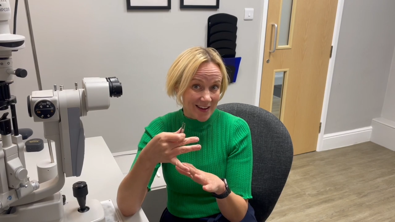
Ms. Rupinder Chahal, MUDr, FRCOphth, is a Consultant Ophthalmologist with a specialist interest in Oculoplastics, at George Eliot NHS Trust.
Dermatochalasis and Ptosis – these two distinct conditions affect the eyelids but what is the difference between them ?
Dermatochalasis
Dermatochalasis refers to excessive skin on the upper or lower eyelid. It may result in a “baggy” or droopy appearance.
- Cause:
- Aging (loss of skin elasticity and weakening of connective tissues).
- Genetic predisposition.
- Trauma or previous eyelid surgery.
- Symptoms:
- Cosmetic concerns (baggy eyelids, tired appearance).
- Obstruction of the superior visual field in severe cases.
- Eyelid heaviness or fatigue.
- Diagnosis:
- Visual field testing, to document obstruction.
- Physical examination – it is possible to pinch the excessive skin. There is an increased measured distance between the upper eyelid margin, and the start of the eyebrow hair: Margin to brow distance is usually much greater than 22mm.
- Treatment:
- Surgical: Blepharoplasty to remove excess skin and/or fat.
Key Feature: Normal eyelid margin position; apparent droopiness is due to redundant skin, not muscle dysfunction.
Ptosis
Drooping of the upper eyelid due to weakness or dysfunction of the any of the muscles responsible for lifting the eyelid:
- Levator palpebrae superioris
- Müller’s muscle.
- Cause:
- Congenital: Maldevelopment of the levator muscle.
- Acquired:
- Aponeurotic – Age-related stretching of the levator tendon
- Neurogenic – e.g. Third nerve palsy, Horner’s syndrome, Myasthaenia Gravis
- Myogenic – e.g. Muscular dystrophies
- Mitochondrial – Kearns-Sayre Syndrome, Chronic Progressive External Ophthalmoplegia (other systemic symptoms will be present)
- Traumatic or mechanical – e.g. Tumour or scar.
- Factors such as prolonged contact lens wear (both RGPs and soft contact lenses), trauma, cataract surgery, or other eye procedures.
- Patient Symptoms:
- Droopy eyelid – one or both sides.
- Obstructed vision if severe.
- Tilting of the head (compensatory chin-up posture) or raising eyebrows to improve vision (frontalis muscle overaction).
- This can lead to discomfort in the eyebrows due to the extra effort required to lift the lids, and a difficulty keeping eyelids open,
- Cosmetic concern
Key Feature: The eyelid margin is lower than normal due to muscle or nerve dysfunction, or levator disinsertion.
What does the Ocular Surface look like in Ptosis Patients?
This will depend on the cause.
- If associated with prolonged soft contact lens or RGP lens wear – dry eyes
- If associated with rubbing eyes:
- Allergy related changes – evert the eyelid and look for chronic changes on the cornea and tarsal conjunctiva.
- Meibomian gland dysfunction with lid margin irritation, or even keratoconus.
- Trauma – look for eyelid scarring on the skin surface, or scarring noted on everting the eyelid.
- Congenital – usually an absent lid crease, with a possible chin up head position
- Clinical Examination:
- Margin reflex distance (MRD) measurement.
- Examination of muscle strength and lid lag.
- Measuring position of the upper lid skin crease.
- Testing for underlying systemic or neurological conditions.
- Checking eye movements, pupil reactions, and assessing optic disc appearance.
Eyelid Measurement Summary Table
Measurement | Definition | Normal Range | How to Perform |
Marginal Reflex Distance (MRD) | Distance between the corneal light reflex and the upper or lower eyelid margin. | MRD1: ≥4–5 mm (upper lid); MRD2: 5 mm (lower lid) | Shine a penlight at the patient’s eyes. Measure the distance from the corneal light reflex to the eyelid margin in millimeters. |
Levator Function | The excursion of the upper eyelid from downgaze to upgaze, measuring the levator muscle’s strength. | 12–15 mm | Firmly place your finger above the eyebrow, to immobilize the brow muscle (Frontalis muscle). Place a ruler next to eyelid margin, ask the patient to look down, then up, and measure the eyelid excursion in millimetres. |
Orbicularis Function | Strength of the orbicularis oculi muscle, responsible for eyelid closure. | Strong, symmetric closure | Ask the patient to close their eyes tightly. Open their eye with your fingers and assess the resistance. Good resistance means good muscle function. |
Skin Crease Measurement | Distance between the eyelid margin and the eyelid crease, indicating levator aponeurosis attachment. | 8–10 mm (males); 9–11 mm (females) | Ask the patient to look down. Measure the distance from the eyelid margin to the visible skin crease. |
Bell’s Phenomenon | Upward and outward rotation of the eyeball during forced eyelid closure. | Present (90% of individuals) | Ask the patient to close their eyes tightly. Gently lift the eyelids and observe if the eyeballs rotate upward and outward. If present, it is reassuring that if there is any lagophthalmos, then the surface is partly protected when asleep, due to this reflex. |
What eye emergencies can manifest with ptosis as one of the symptoms?
3rd nerve Palsy:
This is an emergency, requiring prompt blue light referral to the hospital emergency department.
Signs of third nerve palsy:
- Ptosis
- Diplopia: The affected eye turns outward and downward. (Paralysis of upgaze, downgaze and adduction).
- Dilated pupil: the pupil may be dilated and react sluggishly. If the pupil is affected, it could be a sign of an aneurysm.
- It can be accompanied by a headache, stiff neck, and decreased level of consciousness.
Horners Syndrome:
Horner syndrome may be caused by a stroke, tumour or spinal cord injury.
- Ptosis
- Constriction of pupil
- Decreased sweating on the affected side of the face.
What are the management options for the treatment of Ptosis?
Conservative Treatment Options:
- Artificial tears and lubricating ointments: To alleviate dry eye symptoms and prepare the surface before considering surgery.
- External eyelid crutches: Temporary solutions that hold the eyelid up and reduce visual obstruction.
- Botulinum toxin injections: In select cases, Botulinum Toxin can be used to temporarily relieve mild ptosis,
- Treat underlying conditions: Such as myasthenia gravis or other systemic disorders causing ptosis.
Surgical Options:
Risks
It is important that the patient is counselled about the risks:
- Over or under correction,
- Exacerbation of dry eye,
- Exposure keratopathy,
- Further procedures should any complications occur.
The patient should ideally have an optimised ocular surface prior to ptosis surgery.
Benefits:
- Better lid closure: Surgery corrects the position of the upper eyelid, ensuring that the eye can fully close during blinking. This restores the proper spread of the tear film and prevents dryness.
- Vision improvement: Surgical repair restores peripheral vision blocked by droopy eyelids
Indication | Surgical Objectives | |
Levator Advancement/Resection | Mild to moderate ptosis with good levator function (>8 mm). | Reattachment of a disinserted levator aponeurosis to the tarsal plate, with levator shortening. |
Müller’s Muscle- Conjunctival Resection | Very mild ptosis with excellent levator function. | Resection of Müller’s muscle through the conjunctiva to elevate the eyelid. |
Frontalis Sling Procedure | Severe ptosis with poor levator function (<4 mm). | Uses a sling to connect the eyelid to the frontalis muscle, allowing the brow to lift the lid. |
Post operatively:
Following surgery, the patient will be prescribed:
- Antibiotic ointment for the wound,
- Lubricating ointment to use at night
- Lubricating drops to use during the day
Often there are external skin sutures, which will be removed in clinic approximately two weeks post operatively.
It can take several months to a year to appreciate the outcome. This is due to the stages of tissue healing.
Complications of ptosis surgery
Overcorrection, leading to inadequate lid closure – lagophthalmos.
- This can lead to:
- Dry eyes – an incomplete blink can lead to poor tear film distribution.
- Exposure keratopathy, where the cornea becomes irritated, inflamed and scarred, due to exposure.
- Irritation, burning, and foreign body sensation.
- Corneal damage in more severe cases, particularly if the eyelid is not sufficiently protecting the eye. This can range from dry eye to corneal ulceration. Further surgery/procedures would be indicated.
Mild overcorrection may settle after some time, with eyelid massage.
The Droopy Eyelid – Key Differences Summary:
Dermatochalasis | Ptosis | |
Definition | Excess skin on the eyelids | Drooping eyelid due to muscle/nerve issues |
Cause | Skin laxity (aging, genetics) | Muscle weakness, nerve dysfunction, aging |
Key Feature | Redundant skin above a normally positioned eyelid margin | Low eyelid margin |
Symptoms | “Baggy” eyelids, visual obstruction if severe | Drooping eyelid, obstructed vision, compensatory head posture, brow muscle overaction |
Treatment | Blepharoplasty | Ptosis repair surgery |
Clinical Note
- Dermatochalasis and ptosis can coexist, especially in older adults with age related changes.
- Differentiation is essential for appropriate treatment:
- Dermatochalasis: Focus is on removing excess skin, via blepharoplasty.
- Ptosis: Focus is on repairing the eyelid elevation mechanism.
- Procedures can be combined.
Key Take Home Points:
- Patient Centred Approach: Ask the patient about their primary concern. This helps in creating a sensible management plan tailored to their expectations. Some patients may prefer conservative management and may be satisfied with non-surgical options.
- Health evaluation: Thorough medical history, to ascertain if there is any underlying systemic disease, such as thyroid disease, neurological disorders, or previous trauma.
- Thorough eye examination: Evaluate the ocular surface, tear film, corneal health, and visual fields.
- Manage dry eye disease: Treat underlying ocular surface disorders. Daily blepharitis management if required, and regular lubricants are important. This alone may alleviate symptoms and will also prepare the ocular surface if surgery is indicated.
- Patient expectations: Set realistic goals for functional and cosmetic outcomes.
- Recognise and urgently refer emergency conditions.
Latest Articles


HCP Popup
Are you a healthcare or eye care professional?
The information contained on this website is provided exclusively for healthcare and eye care professionals and is not intended for patients.
Click ‘Yes’ below to confirm that you are a healthcare professional and agree to the terms of use.
If you select ‘No’, you will be redirected to scopeeyecare.com
This will close in 0 seconds