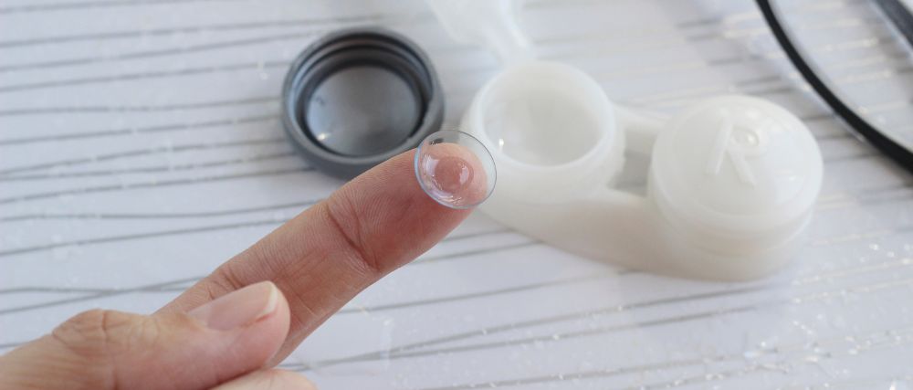
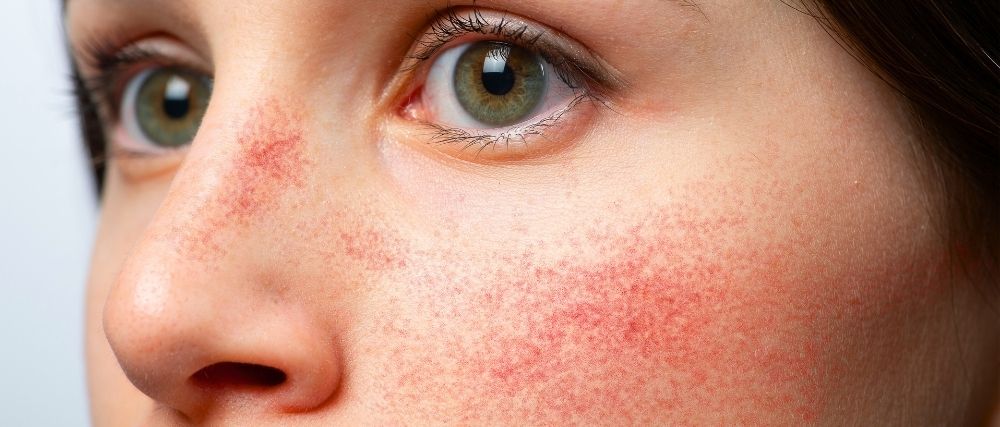
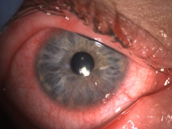
The Role of Ophthalmic Ointments in Night-Time Exposure and RCE: A Roundtable Discussion


Over-the-counter lubricating agents are the go-to for most optometrists when initially addressing dry eye and other ocular surface disorders.1 Typically, this means the use of artificial tears. However, another category of lubricating agents, ophthalmic ointments, should not be forgotten, especially as they are uniquely suited to address several challenging ocular surface conditions – including night-time exposure and recurrent corneal erosion due to properties such as increased contact time. The following is a roundtable discussion between three dry eye and ocular surface disease experts – Drs. Paul Karpecki, Cory Lappin, and Ada Noh – in which they discuss the benefits of ointments, which specific ointments they prefer, how and when they use them, and address questions and concerns regarding their implementation in the management of night-time exposure and recurrent corneal erosion.
Night-Time Exposure
Night-time exposure (NTE) refers to exposure of the cornea and ocular surface to the external environment caused by incomplete closure or an inadequate seal of the eyelids during sleep and is characterized symptomatically as dryness that is worst upon waking.2 This condition is incredibly common but often overlooked. In her clinic, Dr. Noh notes that up to 85% of her patients have some form of a nocturnal eyelid seal issue, oftentimes without any obvious clinical signs, which is a relatively common occurrence.2 When discussing if the high prevalence of night-time exposure related issues is increasing or just becoming more commonly recognized, the group agrees that NTE has likely always been an issue but is receiving more attention due to increased awareness around dry eye and ocular surface-related conditions in general. Dr. Lappin also sees a high prevalence of NTE in his clinic with “about one third of patients having signs of NTE and a much higher percentage experiencing some form of nocturnal exposure issue without any overt associated signs.”
A major reason NTE is so prevalent is due to the fact that it can stem from numerous conditions. It can be caused by “classic” lagophthalmos in which the lids do not physically come together,3,4 an inadequate eyelid seal in which the upper and lower lids touch but do not completely seal,2 excessive lid laxity due to the loss of skin rigidity from aging or floppy eyelid syndrome associated with obstructive sleep apnea,4-8 inadequate closure following blepharoplasty,3,4 or pathological conditions that impact the ability of the lids to close like Bell’s palsy.3,4 Accordingly, Dr. Lappin puts the various forms of NTE “in different ‘buckets,’ such as true lagophthalmos, poor lid seal, surgically induced exposure, and pathology-related cases.” Of these various causes of NTE, he finds that a poor lid seal is by far the most common, which is a sentiment echoed by Drs. Noh and Karpecki. Additionally, inferiorly located corneal staining is an indication of exposure keratitis with the group noting this tends to occur when the exposure is chronic and potentially represents a more progressed form of the condition. “I tend to treat staining as a body’s cry for help,” says Dr. Noh of such cases.
In the context of dry eye disease, where does NTE fit? “That’s a complicated question because it depends on the patient as NTE and dry eye can fall under the same umbrella of related conditions,” explains Dr. Noh. Dr. Lappin agrees, “Dry eye and NTE can be distinct conditions, however they can also overlap and impact one another, so it is really a two-way street.” Dr. Karpecki also emphasizes the clinical importance of recognizing nocturnal exposure as a related but still independent entity, especially in presumed dry eye patients that are unresponsive to traditional dry eye therapies because their symptoms may actually be caused by NTE. In fact, one study showed that refractory dry eye patients were four times more likely to have a compromised lid seal than asymptomatic patients.9 “I think this is the number one cause of nonresponsive dry eye,” explains Dr. Karpecki, and in some cases NTE “is their entire problem.” Dr. Lappin finds this to be consistent with his clinical experience as well, noting some patients report complete or near complete resolution in their symptoms after addressing NTE alone. However, other patients may have both NTE and dry eye, in which a poor-quality tear film fails to protect their ocular surface, worsening their nocturnal exposure, and, in turn, further disrupting their tear film creating a vicious cycle.3,4,9,10
Given the significant impact NTE can have on the ocular surface, how is it diagnosed and differentiated from other ocular surface conditions? For Dr. Noh, it all starts with patient history, “I take history myself on purpose because I find out so much information,” further explaining that the number one way she diagnoses NTE is from patient history and simply asking if they experience symptoms upon waking. In addition to asking about patient history and symptoms, Dr. Karpecki states he “likes confirmatory tests,” such as the Korb-Blackie light test (where a transilluminator is placed against the upper lid of a patient’s closed eye to see if any light shines out between the lids which indicates areas of an inadequate seal),2 but morning symptoms alone can validate the presence of NTE or inadequate lid seal (ILS). “Just by using a quick test to look at the eyelids closely and knowing the history, will give you a good indication you are dealing with this condition.” Dr. Lappin takes a similar approach to diagnosing NTE, asking patients if they have symptoms upon waking and then checking for signs of incomplete lid closure by having the patient tilt their head back and using a transilluminator to look for lagophthalmos or a poor lid seal.
Now that NTE has been diagnosed, how is it treated? According to the group, ointments should be top of mind when managing NTE.3,4,8 “The time to start thinking about ointments is when you can use them overnight,” states Dr. Karpecki. Dr. Lappin agrees, stating “The number one condition that comes to mind in terms of ointment use is night-time exposure.” He further explains that he takes a systematic, three step-approach when treating NTE, starting with the use of ointment before bed as this tends to be the easiest and least cumbersome initial treatment, followed by the use of moisture chamber sleep goggles as the next step if needed, and finally the use of lid taping or adhesive lid seals if issues persist. However, he still recommends the use of ointment along with sleep goggles or sleep strips.
Dr. Noh states that she approaches NTE the exact same way and emphasizes the importance of patient education, “Another thing that I think is important to explain to patients is that this is an anatomical issue.” She goes on to explain that this is particularly useful in helping patients understand why long-term treatment is needed. The regular use of treatment, such as topical ointment, is necessary to keep the eyes protected because the lids are still not closing properly. She also finds the use of analogies helpful with patient education, “It’s like using Saran wrap on your leaky windows in the winter, the window is still leaky but you are trying to give it some extra protection.”
One of the challenges of managing NTE is that treatments can be blurring or obstruct vision, which can prompt concern from patients. Dr. Noh addresses such concerns by having the patient alternate the use of ointment in one eye each night, so if they need to get up to use the restroom or they have a baby, they will still be able to see. Dr. Karpecki, who also uses a mix of ointment and lid seals to manage NTE, takes a similar approach, “If I’m going to use a night seal strip, I’m typically going to have them start with only one eye because many patients need to get up in the middle of the night and they need to get used to it, so I’ll have them use ointment in the other eye, and then I’ll have them alternate eyes the next night.”
Recurrent Corneal Erosion (RCE)
Another relatively common condition that has a nocturnal element is recurrent corneal erosion (RCE).
In cases of RCE, the attachment of the corneal epithelium to the basement membrane is compromised, typically due to traumatic injury, a structural abnormality like epithelial basement membrane dystrophy (EBMD), or even NTE.11-15 This poor adhesion can be further weakened by changes to the ocular surface environment during sleep, resulting in repeated episodes of the fragile epithelium being ripped away by shearing forces of the lid, usually upon waking.11,13 The characteristic symptoms are sharp, often intense, pain owing to the dense innervation of the cornea that is forty-times more innervated than tooth pulp.11-18
Management of RCE can be complex and varies from doctor to doctor, however Drs. Karpecki, Lappin, and Noh all agree that treatment of RCE has two phases – acute, when there is an active abrasion or erosion, and long-term, when the goal is to prevent further episodes. This follows the natural progression of corneal wound healing as the cornea re-epithelializes relatively quickly, typically within 24 hours, but it can take up to six weeks for the hemidesmosomes to form and fully anchor the epithelium to the basement membrane.14,19-21 Accordingly, Dr. Noh addresses the acute stage with a bandage contact lens or amniotic membrane as needed, as will Dr. Karpecki who chooses his treatment strategy based on severity, often debriding loose epithelium and using an amniotic membrane in recalcitrant cases. He also notes there are two components that need to be addressed when managing RCE – epithelial edema that occurs overnight and proinflammatory MMPs, both of which can prevent the healing and adherence of the epithelium to the basement membrane, and can be managed with hyperosmotic and anti-inflammatory agents, respectively.11,14 Dr. Lappin also approaches treatment with these factors in mind, using serial bandage contact lenses to provide a protective barrier for the fragile, healing epithelium along with low dose oral doxycycline and a topical steroid to inhibit the actions of MMPs.11,14,22,23
Once the initial acute phase of RCE has been managed, long-term management aimed at preventing future episodes becomes crucial,11-14 as Dr. Noh states “We know once a patient has had a recurrent corneal erosion, they are more susceptible to have it happen again, so we have to provide something to protect the cornea through the night-time.” This long-term management of RCE “is where ointment becomes key” according to Dr. Karpecki. Because episodes of RCE typically occur upon waking due to changes to the ocular surface environment during sleep, Dr. Lappin approaches long-term management similarly to NTE treatment, recommending the use of nighttime ointment as “It can be a good way to reduce friction and the sheering forces of the lids especially upon waking.”
OPTASE HYLO Night
When it comes to managing nocturnal conditions like NTE and RCE, Dr. Karpecki states “Ointments are an underutilized option that can be far superior in conditions like this compared to other things we could use,” explaining that “they have a longer contact time” on the ocular surface compared to traditional drops which helps protect the cornea throughout the night.24-28 So, in cases where the use of ointment is beneficial, is there a specific ointment that is recommended? Dr. Karpecki explains this is situational, as he may use a hyperosmotic ointment during the acute phase of RCE treatment to help reduce edema but notes patients typically don’t like to stay on hyperosmotic ointments because they can be uncomfortable and are not necessary after a couple of months. This is when he specifically prefers to use an ointment like OPTASE HYLO Night (SCOPE Health Inc) for maintenance as it is better suited to “reduce friction and desiccation,” which he will even recommend up to three times per day if needed.
Dr. Lappin explains that he “didn’t routinely use ointments prior to the availability of “OPTASE HYLO Night” primarily because many traditional bland and antibiotic ointments contain preservatives, patients sometimes find them difficult to use, and he has not found them to be as effective as he desires. However, he finds OPTASE HYLO Night easier for patients to use and more effective clinically, which he attributes to its formulation. Dr. Noh also specifically recommends OPTASE HYLO Night to her patients, highlighting that its preservative-free formulation “is huge for me” as preservative exposure can be detrimental to the ocular surface.29-32 A preservative-free formulation is especially important for ophthalmic ointments given their longer contact times24-27 compared to other drops, as ointments containing preservatives can expose the ocular surface to these irritating agents for a longer period of time.
What sets OPTASE HYLO Night apart from other ointments? “Not all ointments are the same,” Dr. Lappin explains and points to OPTASE HYLO Night’s unique formulation as a major reason for its efficacy. Traditional ointments are primarily composed of petroleum and little else, whereas OPTASE HYLO Night contains white petrolatum which acts as a lubricant, light liquid paraffin which increases water retention and reduces evaporation, vitamin A in the form of retinol palmitate, which is a lipophilic oil that readily mixes with the other ingredients giving the ointment its “soft” feeling and easy flow, as well as lanolin, which reduces the melting point of the ointment which can cut down on the blurring effect and “tackiness” commonly associated with ointments.33-36 Topical vitamin A has also been shown to promote corneal and conjunctival wound healing37,38 and improve signs and symptoms of dry eye.38,39
How do you approach some of the challenges associated with ointment use?
The use of ointment poses a few potential challenges, with the blurring effect typically the top consideration.24-28 Additionally, one of the benefits of using ointments before bed is that the blurring effect will wear off overnight, as Dr. Karpecki explains, “if the patient experiences some blur it is not a major issue because they are sleeping and by the time they wake up most of, if not all, the blur will have dissipated.” One study that investigated the impact of ointment-related blur even noted that “eye ointment is still an essential treatment modality in clinical practice” despite these blurring effects.28 However, these are concerns that should be addressed when recommending ointments to patients, as Dr. Lappin noted that “many patients are concerned their vision will be blurred for hours even when they wake up, and they will have a buildup of ‘gunk’ along with their lids and lashes being stuck together.” However, all three doctors have found that these issues are not as common with OPTASE HYLO Night. In Dr. Noh’s experience “patients like HYLO Night more, they find it is easier to use and don’t mind these effects compared to other ointments.” Because of OPTASE HYLO Night’s specific formulation, once the ointment is on the eye, the body’s heat will cause it to melt33-35,40 “giving it a more gel-like consistency compared to the traditional Vaseline-like feel experienced with conventional ointments,” adds Dr. Lappin. This difference was also shown in a small, open-label study that looked at the ease of use, tolerability, and efficacy of OPTASE HYLO Night compared to a traditional ointment containing petrolatum and mineral oil. The results of the study found 82% of patients in the HYLO Night arm continued use of the ointment, whereas 82% of patients in the traditional ointment arm discontinued use, noting blurred vision as the most common reason for discontinuation as well as difficulty with instillation.41 This study reinforces the superior tolerability and ease of use of OPTASE HYLO Night that has been observed by all three doctors clinically.
Drs. Karpecki, Lappin, and Noh all also emphasize the importance of giving patients a specific recommendation. OPTASE HYLO Night is the only ointment Dr. Noh carries in her office so she knows her patients are getting exactly what they need. Likewise, Dr. Lappin carries OPTASE HYLO Night for the same reason, and states that even if you don’t stock dry eye products in your office “it is crucial to tell your patients exactly what you want them to use, because if you don’t, they will inevitably pick the cheapest option they can find” which is more likely to have an older formulation and contain harsh preservatives.
Another aspect of successful ointment use is education regarding its instillation, explains Dr. Noh. “I tell the patient to pull down on the lower lid and make a pocket and to put the ointment in the pocket.” This can be particularly helpful as many patients assume they have to get the ointment directly onto the eye when instilling it. However, Dr. Noh also recommends explicitly telling patients the ointment does actually need to go into their eyes, as some may mistakenly think the ointment goes on their eyelids given that most of their previous experiences with ointments have likely been for use on their skin. Dr. Lappin adds that “a little bit goes a long way,” and that they only need to use a small amount of OPTASE HYLO Night, not a whole strip or ribbon, as they will still get the benefits of the ointment while reducing some of the side effects, like blur.24
Are there any concerns regarding the use of ointment?
A previous study found that symptoms of RCE actually worsened after patients used bland ointment for two months following their initial injury.42 For this reason, the use of bland ointments for RCE has been cautioned against. However, as Drs. Karpecki, Lappin, and Noh note this does not mean that the use of ointments should be avoided entirely, but rather that specific ointments should be used in the appropriate context. For instance, this worsening of healing was observed when ointment was used in the acute phase of RCE, whereas the roundtable recommends using ointment after this initial period as a form of longer-term maintenance. Additionally, ointment composition also plays a large role as older, waxier petroleum-based ointments that are commonly referred to as “bland ointments” have been shown to interfere with wound healing, however more contemporary formulations have not shown any negative impacts on healing.43 This highlights the importance of using modern ointment formulations, like OPTASE HYLO Night, in the management of ocular surface disease as they are designed to more effectively and safely interact with the ocular surface.
Pull-out Box about Vitamin A:
One of the ingredients that differentiates OPTASE HYLO Night from other ophthalmic ointments is vitamin A in the form of retinol palmitate.33 One potential concern many optometrists have is the relationship between vitamin A and meibomian gland toxicity.44-50 In this context, it is crucial to understand that not all forms of vitamin A are the same or have the same effects on the ocular surface.51 Retinoids such as isotretinoin and tretinoin can be damaging to the meibomian glands, especially in oral and dermatological formulations.44-50 However, studies have shown topical retinol palmitate not only to be safe,39 but beneficial to the ocular surface as topical forms have enhanced corneal and conjunctival wound healing,37,38 improved mucin production and goblet cell health,37,38,52-55 and improved signs and symptoms of dryness.39,38 In fact, vitamin A is so essential to ocular surface homeostasis that it is stored in and secreted from the lacrimal gland,56,57 as it is vital to cellular differentiation and proliferation as well as epithelial cell and mucus membrane health.58 Consequently, vitamin A deficiency can result in keratinization of the ocular surface, squamous metaplasia of the cornea and conjunctiva, and a decrease in goblet cells which can result in mucin deficiency.48,59,60
Therefore, while it is imperative to help patients avoid potentially harmful vitamin A derivatives, optometrists should be equally aware of the forms of vitamin A that are both beneficial and essential to ocular surface health.
References
- Semp DA, Beeson D, Sheppard AL, Dutta D, Wolffsohn JS. Artificial Tears: A Systematic Review. Clin Optom (Auckl). 2023;15:9-27. Published 2023 Jan 10. doi:10.2147/OPTO.S350185
- Blackie CA, Korb DR. A novel lid seal evaluation: the Korb-Blackie light test. Eye Contact Lens. 2015;41(2):98-100. doi:10.1097/ICL.0000000000000072
- Latkany RL, Lock B, Speaker M. Nocturnal lagophthalmos: an overview and classification. Ocul Surf. 2006;4(1):44-53. doi:10.1016/s1542-0124(12)70263-x
- Fu L, Patel BC. Lagophthalmos. [Updated 2023 Jul 24]. In: StatPearls [Internet]. Treasure Island (FL): StatPearls Publishing; 2024 Jan-. Available from: https://www.ncbi.nlm.nih.gov/books/NBK560661/
- Chhadva P, McClellan AL, Alabiad CR, Feuer WJ, Batawi H, Galor A. Impact of Eyelid Laxity on Symptoms and Signs of Dry Eye Disease. Cornea. 2016;35(4):531-535. doi:10.1097/ICO.0000000000000786
- De Gregorio A, Cerini A, Scala A, Lambiase A, Pedrotti E, Morselli S. Floppy eyelid, an under-diagnosed syndrome: a review of demographics, pathogenesis, and treatment. Ther Adv Ophthalmol. 2021;13:25158414211059247. Published 2021 Dec 5. doi:10.1177/25158414211059247
- Scarabosio A, Surico PL, Patanè L, et al. The Overlooked Floppy Eyelid Syndrome: From Diagnosis to Medical and Surgical Management. Diagnostics (Basel). 2024;14(16):1828. Published 2024 Aug 21. doi:10.3390/diagnostics14161828
- Mastrota KM. Impact of floppy eyelid syndrome in ocular surface and dry eye disease. Optom Vis Sci. 2008;85(9):814-816. doi:10.1097/OPX.0b013e3181852777
- Korb D, Blackie C, Nau A. Prevalence of compromised lid seal in symptomatic refractory dry eye patients and asymptomatic patients. Invest Ophthalmol Vis Sci. 2017; 58:2696.
- Liu DT, Di Pascuale MA, Sawai J, Gao YY, Tseng SC. Tear film dynamics in floppy eyelid syndrome. Invest Ophthalmol Vis Sci. 2005;46(4):1188-1194. doi:10.1167/iovs.04-0913
- Miller DD, Hasan SA, Simmons NL, Stewart MW. Recurrent corneal erosion: a comprehensive review. Clin Ophthalmol. 2019;13:325-335. Published 2019 Feb 11. doi:10.2147/OPTH.S157430
- Hykin PG, Foss AE, Pavesio C, Dart JK. The natural history and management of recurrent corneal erosion: a prospective randomised trial. Eye (Lond). 1994;8 ( Pt 1):35-40. doi:10.1038/eye.1994.6
- Ramamurthi S, Rahman MQ, Dutton GN, Ramaesh K. Pathogenesis, clinical features and management of recurrent corneal erosions. Eye (Lond). 2006;20(6):635-644. doi:10.1038/sj.eye.6702005
- Das S, Seitz B. Recurrent corneal erosion syndrome. Surv Ophthalmol. 2008;53(1):3-15. doi:10.1016/j.survophthal.2007.10.011
- Sturrock GD. Nocturnal lagophthalmos and recurrent erosion. Br J Ophthalmol. 1976;60(2):97-103. doi:10.1136/bjo.60.2.97
- Eguchi H, Hiura A, Nakagawa H, Kusaka S, Shimomura Y. Corneal Nerve Fiber Structure, Its Role in Corneal Function, and Its Changes in Corneal Diseases. Biomed Res Int. 2017;2017:3242649. doi:10.1155/2017/3242649
- Müller LJ, Marfurt CF, Kruse F, Tervo TM. Corneal nerves: structure, contents and function. Exp Eye Res. 2003;76(5):521-542. doi:10.1016/s0014-4835(03)00050-2
- Rózsa AJ, Beuerman RW. Density and organization of free nerve endings in the corneal epithelium of the rabbit. Pain. 1982;14(2):105-120. doi:10.1016/0304-3959(82)90092-6
- Hamill MB. Mechanical Injury. In: Krachmer JH, Mannis MJ, Holland EJ eds. Cornea. London: Elsevier; 2011:1088-1105.
- Remington LA. The Cornea and Sclera. In: Clinical Anatomy of the Visual System. Newton, Mass.: Butterworth-Heinemann; 1998:9-29.
- Binder, PS, Wickham GM, Zavala EY, et al. Corneal Anatomy and Wound Healing. In: Barraquer, JI, Binder PS, Buxton JN (eds.). Symposium on Medical and Surgical Diseases of the Cornea. St. Louis: CV Mosby Company; 1980:1-35.
- Dursun D, Kim MC, Solomon A, Pflugfelder SC. Treatment of recalcitrant recurrent corneal erosions with inhibitors of matrix metalloproteinase-9, doxycycline and corticosteroids. Am J Ophthalmol. 2001;132(1):8-13. doi:10.1016/s0002-9394(01)00913-8
- Fraunfelder FW, Cabezas M. Treatment of recurrent corneal erosion by extended-wear bandage contact lens. Cornea. 2011;30(2):164-166. doi:10.1097/ICO.0b013e3181e84689
- Robin JS, Ellis PP. Ophthalmic ointments. Surv Ophthalmol. 1978;22(5):335-340. doi:10.1016/0039-6257(78)90178-9
- Greaves JL, Wilson CG, Birmingham AT. Assessment of the precorneal residence of an ophthalmic ointment in healthy subjects. Br J Clin Pharmacol. 1993;35(2):188-192.
- Bao Q, Burgess DJ. Perspectives on Physicochemical and In Vitro Profiling of Ophthalmic Ointments. Pharm Res. 2018;35(12):234. Published 2018 Oct 15. doi:10.1007/s11095-018-2513-3
- Baranowski P, Karolewicz B, Gajda M, Pluta J. Ophthalmic drug dosage forms: characterisation and research methods. ScientificWorldJournal. 2014;2014:861904. Published 2014 Mar 18. doi:10.1155/2014/861904
- Hiraoka T, Yamamoto T, Okamoto F, Oshika T. Time course of changes in ocular wavefront aberration after administration of eye ointment. Eye (Lond). 2012;26(10):1310-1317. doi:10.1038/eye.2012.142
- HYLO Night® A preservative free solution to manage night-time dry eye. Scope Eyecare. Accessed December 17, 2024. https://scopeeyecare.com/product/hylo-night/.
- Kahook MY, Rapuano CJ, Messmer EM, Radcliffe NM, Galor A, Baudouin C. Preservatives and ocular surface disease: A review. Ocul Surf. 2024;34:213-224. doi:10.1016/j.jtos.2024.08.001
- Baudouin C, Labbé A, Liang H, Pauly A, Brignole-Baudouin F. Preservatives in eyedrops: the good, the bad and the ugly. Prog Retin Eye Res. 2010;29(4):312-334. doi:10.1016/j.preteyeres.2010.03.001
- Kaur IP, Lal S, Rana C, Kakkar S, Singh H. Ocular preservatives: associated risks and newer options. Cutan Ocul Toxicol. 2009;28(3):93-103. doi:10.1080/15569520902995834
- Optase Hylo Night Eye Ointment. U.S. National Library of Medicine. November 2023. Accessed December 17, 2024. https://dailymed.nlm.nih.gov/dailymed/fda/fdaDrugXsl.cfm?setid=c1991caa-51b9-13d1-e053-2995a90a1293.
- Fiscella R, Jensen M. Ophthalmic disorders. In: Krinsky D, Berardi R, Ferreri S, et al, eds. Handbook of Nonprescription Drugs. 18th ed. Washington, DC: American Pharmacists Association; 2015.
- Terrie YC. Relieving Dry Eye: What Every Patient Needs to Know. Pharmacy Times. May 12, 2016. Accessed December 17, 2024. https://www.pharmacytimes.com/view/relieving-dry-eye-what-every-patient-needs-to-know.
- Ursapharm 2024. Data on File.
- Toshida H, Odaka A, Koike D, Murakami A. Effect of retinol palmitate eye drops on experimental keratoconjunctival epithelial damage induced by n-heptanol in rabbit. Curr Eye Res. 2008;33(1):13-18. doi:10.1080/02713680701827696
- Hyon JY, Han SB. Dry Eye Disease and Vitamins: A Narrative Literature Review. Applied Sciences. 2022; 12(9):4567. https://doi.org/10.3390/app12094567
- Toshida H, Funaki T, Ono K, et al. Efficacy and safety of retinol palmitate ophthalmic solution in the treatment of dry eye: a Japanese Phase II clinical trial [published correction appears in Drug Des Devel Ther. 2021 Feb 24;15:813-816. doi: 10.2147/DDDT.S301482]. Drug Des Devel Ther. 2017;11:1871-1879. Published 2017 Jun 23. doi:10.2147/DDDT.S137825
- Park EK, Song KW. Rheological evaluation of petroleum jelly as a base material in ointment and cream formulations: steady shear flow behavior. Arch Pharm Res. 2010;33(1):141-150. doi:10.1007/s12272-010-2236-4
- El-khashab T, Quah SA, Chiam P, Rudy-Fitzgerald L, Noden D. Comparison of the clinical efficacy of HYLO-Night® and Lacri-Lube® in patients with dysfunction tear syndrome. MBA Eye Care Centre, Leighton Hospital, Crewe, Cheshire, CW 4QJ, UK. 2024.
- Eke T, Morrison DA, Austin DJ. Recurrent symptoms following traumatic corneal abrasion: prevalence, severity, and the effect of a simple regimen of prophylaxis. Eye (Lond). 1999;13 ( Pt 3a):345-347. doi:10.1038/eye.1999.87
- Hanna C, Fraunfelder FT, Cable M, Hardberger RE. The effect of ophthalmic ointments on corneal wound healing. Am J Ophthalmol. 1973;76(2):193-200. doi:10.1016/0002-9394(73)90159-1
- Moy A, McNamara NA, Lin MC. Effects of Isotretinoin on Meibomian Glands. Optom Vis Sci. 2015;92(9):925-930. doi:10.1097/OPX.0000000000000656
- Zhang P, Tian L, Bao J, et al. Isotretinoin Impairs the Secretory Function of Meibomian Gland Via the PPARγ Signaling Pathway. Invest Ophthalmol Vis Sci. 2022;63(3):29. doi:10.1167/iovs.63.3.29
- Ding J, Kam WR, Dieckow J, Sullivan DA. The influence of 13-cis retinoic acid on human meibomian gland epithelial cells. Invest Ophthalmol Vis Sci. 2013;54(6):4341-4350. Published 2013 Jun 26. doi:10.1167/iovs.13-11863
- Zakrzewska A, Wiącek MP, Słuczanowska-Głąbowska S, Safranow K, Machalińska A. The Effect of Oral Isotretinoin Therapy on Meibomian Gland Characteristics in Patients with Acne Vulgaris. Ophthalmol Ther. 2023;12(4):2187-2197. doi:10.1007/s40123-023-00737-6
- Samarawickrama C, Chew S, Watson S. Retinoic acid and the ocular surface. Surv Ophthalmol. 2015;60(3):183-195. doi:10.1016/j.survophthal.2014.10.001
- Knop E, Knop N, Millar T, Obata H, Sullivan DA. The international workshop on meibomian gland dysfunction: report of the subcommittee on anatomy, physiology, and pathophysiology of the meibomian gland. Invest Ophthalmol Vis Sci. 2011;52(4):1938-1978. Published 2011 Mar 30. doi:10.1167/iovs.10-6997c
- Aslan Bayhan S, Bayhan HA, Çölgeçen E, Gürdal C. Effects of topical acne treatment on the ocular surface in patients with acne vulgaris. Cont Lens Anterior Eye. 2016;39(6):431-434. doi:10.1016/j.clae.2016.06.009
- Fogagnolo P, De Cilla’ S, Alkabes M, Sabella P, Rossetti L. A Review of Topical and Systemic Vitamin Supplementation in Ocular Surface Diseases. Nutrients. 2021;13(6):1998. Published 2021 Jun 10. doi:10.3390/nu13061998
- Toshida H, Tabuchi N, Koike D, et al. The effects of vitamin A compounds on hyaluronic acid released from cultured rabbit corneal epithelial cells and keratocytes. J Nutr Sci Vitaminol (Tokyo). 2012;58(4):223-229. doi:10.3177/jnsv.58.223
- Tabuchi N, Toshida H, Koike D, et al. Effect of Retinol Palmitate on Corneal and Conjunctival Mucin Gene Expression in a Rat Dry Eye Model After Injury. J Ocul Pharmacol Ther. 2017;33(1):24-33. doi:10.1089/jop.2015.0161
- Tseng SC. Topical tretinoin treatment for severe dry-eye disorders. J Am Acad Dermatol. 1986;15(4 Pt 2):860-866. doi:10.1016/s0190-9622(86)70243-0
- Kobayashi TK, Tsubota K, Takamura E, Sawa M, Ohashi Y, Usui M. Effect of retinol palmitate as a treatment for dry eye: a cytological evaluation. Ophthalmologica. 1997;211(6):358-361. doi:10.1159/000310829
- Ubels JL, MacRae SM. Vitamin A is present as retinol in the tears of humans and rabbits. Curr Eye Res. 1984;3(6):815-822. doi:10.3109/02713688409000793
- Ubels JL, Osgood TB, Foley KM. Vitamin A is stored as fatty acyl esters of retinol in the lacrimal gland. Curr Eye Res. 1988;7(10):1009-1016. doi:10.3109/02713688809015147
- McEldrew EP, Lopez MJ, Milstein H. Vitamin A. [Updated 2023 Jul 10]. In: StatPearls [Internet]. Treasure Island (FL): StatPearls Publishing; 2024 Jan-. Available from: https://www.ncbi.nlm.nih.gov/books/NBK482362/
- Sommer Effects of vitamin A deficiency on the ocular surface. Ophthalmology. 1983;90(6):592-600. doi:10.1016/s0161-6420(83)34512-7
- Davidson HJ, Kuonen VJ. The tear film and ocular mucins. Vet Ophthalmol. 2004;7(2):71-77. doi:10.1111/j.1463-5224.2004.00325.x
Latest Articles


HCP Popup
Are you a healthcare or eye care professional?
The information contained on this website is provided exclusively for healthcare and eye care professionals and is not intended for patients.
Click ‘Yes’ below to confirm that you are a healthcare professional and agree to the terms of use.
If you select ‘No’, you will be redirected to scopeeyecare.com
This will close in 0 seconds