Introduction
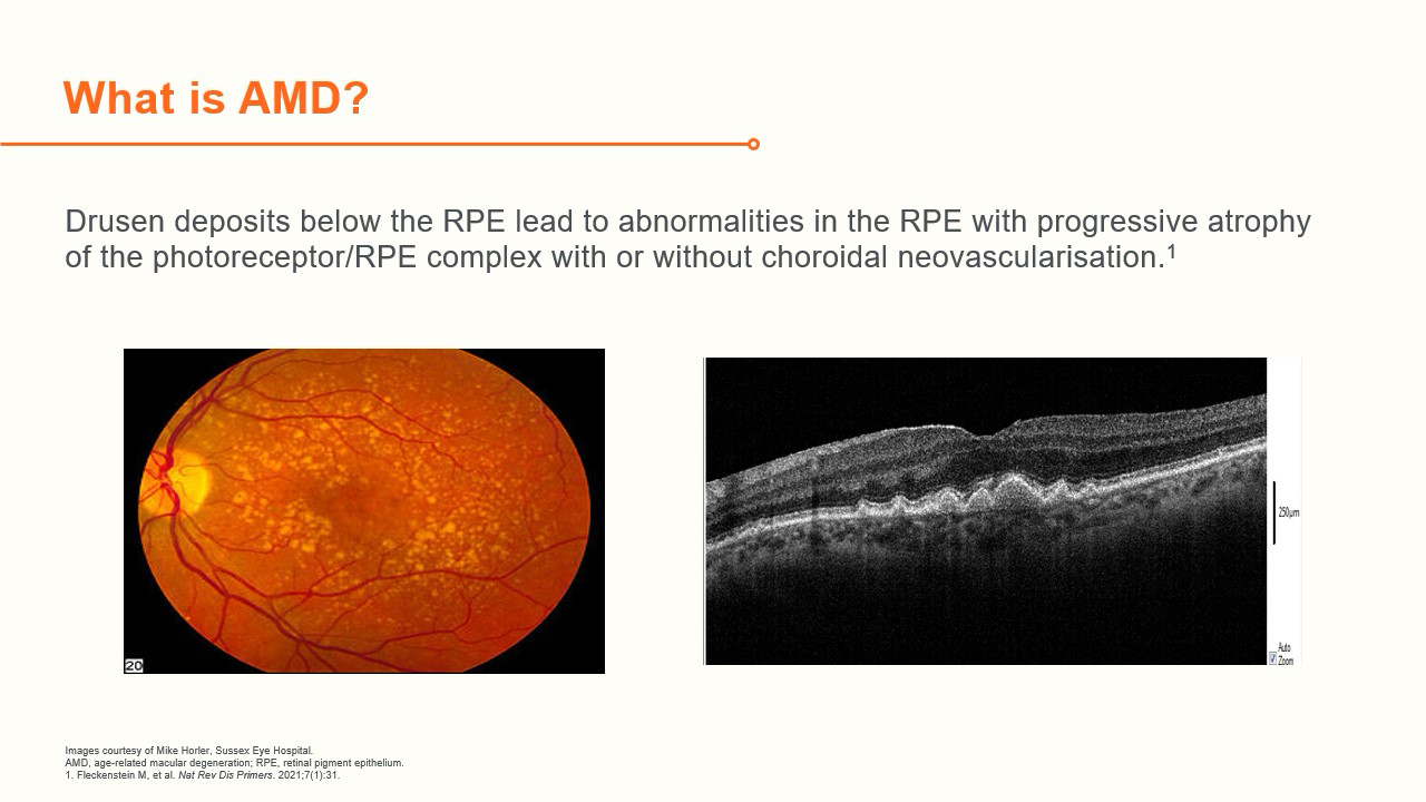
Age-related Macular Degeneration (AMD) is a disease which affects the macula and is defined as a maculopathy which affects people over the age of 50 and includes the presence of drusen and altered retinal pigment epithelium (RPE) pigmentation. Drusen are extracellular deposits of lipids, proteins, and cellular debris which are found within the layers of the retina and appear as small, yellow deposits on dilated eye exams.
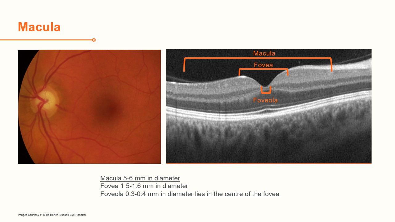
The macula is the central region of the retina and is responsible for fine detailed vision and colour vision. AMD is part of the aging process, where metabolic waste products from the retina build up underneath the retina. These are normally removed in younger patients but accumulate in older patients and damages the sensitive retina at the macula. This leads to a slow and progressive thinning and degeneration of the photoreceptors and the RPE gradually leads to failure of central vision. The larger the drusen, the greater the area and the larger the areas of RPE change, the greater the risk of late AMD.
AMD is the leading cause of blindness in western countries and is responsible for two thirds of all sight impairment registrations in the UK. In 2006 it was estimated that 30-50 million people world-wide were affected, with the incidence increasing with age.1 More recent studies show that the world-wide prevalence of AMD is expected to increase from 198 million in 2020 to 288 million in 2040.2 There are 2 main types of AMD: Dry (non-exudative, non-neovascular) AMD and Wet (exudative, neovascular) AMD. Later stages of the disease are associated with impairment of vision.
Dry (non-exudative, non-neovascular) AMD2
- Most common form (80%)
- Slower progression, usually bilateral
- Geographic atrophy (GA) is the advanced stage of dry AMD
Wet (exudative, neovascular) AMD
- Less common than dry
- More rapid progression to advanced sight loss
- Main manifestations are choroidal neovascular membrane (CNVM) and pigment epithelium detachments (PED)
Approximately 90% of people with dry AMD will only have mild or moderate gradual visual loss and lifestyle measures (not smoking, taking a diet rich in dark green leafy vegetables, Omega 3, etc.) can slow its progression.3 Although it is the less aggressive form, advanced late Dry AMD still causes about 20-25% of AMD related severe visual loss.
Wet (neovascular) AMD is caused by the growth of new vessels from the choroidal circulation. Hypoxia in the RPE leads to Vascular Endothelial Growth Factor (VEGF) being released, which causes new vessels to grow, in order to try and stabilize the nutrition and support of the RPE by the choroid. These vessels can leak or bleed, which causes disruption and damage to the macula through:
- Sub-RPE fluid (Serous Pigment epithelium detachment)
- Sub-retinal fluid
- Intra-retinal fluid
- Sub-retinal Hyperreflective material (SHRM)
- Haemorrhages
The fluid usually causes distortion and rapid visual loss. In some early cases the vision can be preserved. The most extreme vision loss is usually due to large haemorrhages under the macula (sub macula haemorrhage). Wet AMD affects about 10-20% of those who have AMD. It is important to recognise it early as it is potentially treatable. However, if untreated it causes severe visual loss it is responsible for 90% of severe visual acuity loss from AMD.4
AMD has a profound effect on patient quality of life. A patient clinical study looking at dry AMD and its impact found most dry AMD patients suffer with difficulty driving, reading, completing activities of daily living, frustration, dependency on other people, stress and anxiety and lack of confidence.2